Anatomy and Histology of the Hippocampus
Section Reference: Cliques and cavities in the human connectome by Ann E. Sizemore, Chad Giusti, Ari Kahn, Jean M. Vettel, Richard F. Betzel, and Danielle S. Bassett \(^{[1]}\)
Introduction
The hippocampus is distinguished externally as a layer of densely packed neurons that form a S-shaped structure and extends to the temporal lobe of the cerebral cortex. It is also a sub-cortical structure in the limbic lobe, and contains two parts: cornu ammonis CA and dentate gyrus DG, where the hippocampal sulcus separates both parts. The parts curve into each other and below the sulcus lies the subiculum. Since the hippocampus is a part of the allocortex or archicortex, there exists a zone that separates the hippocampus from the neocortex. The hippocampal set is the entire set of hippocampal formation. This formation is comprised by some anatomists into four main sections: Hippocampus proper, dentate gyrus, subiculum, and the entorhinal area EC. This entire set of hippocampus zones is called the hippocampal formation. The hippocampus is supplied blood by the posterior Cerebral artery and is covered by the choroid plexus.
Furthermore, the hippocampus lies posterior to the limbic system, while the anterior is the amygdala. In adult humans, the volume of the hippocampus in each hemisphere is \(\sim 3-3.5 \mathrm{~cm}^3\), where, relative to the volume of the neocortex \(\sim 320-420 \mathrm{~cm}^3\) is \(100\) times smaller compared to the cerebral cortex.
The general function of the hippocampus is organizing emotional responses, which are expressed in the cingulate gyrus via mammillary bodies. Further functions included recollecting experience and imagining the future, playing a role in learning, memory, and spatial navigation.
Cornu Ammonis
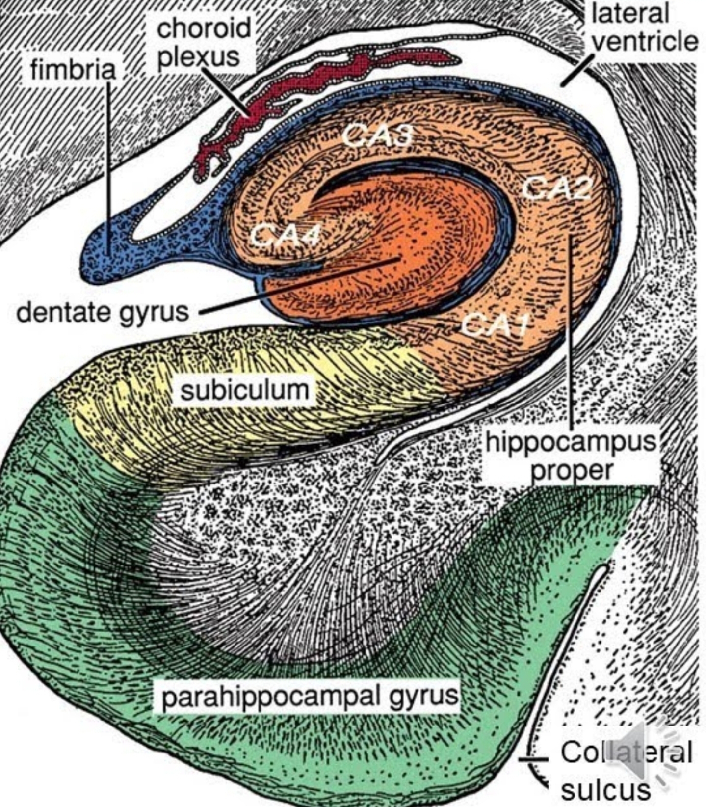
Image Source: Neurosurgery written board crash course - Hippocampus \(^{[2]}\)
Anatomy and Histology
Histology tells us that the hippocampus proper or Cornus Ammonis is divided into four histological domains: CA1, CA2, CA3, and CA4. Across from the CA1 is the subiculum, which is a formation connecting the hippocampus to the entorhinal cortex in the ventricle. Where the entorhinal cortex inputs to the dentata gyrus and plays an important role in pattern recognition and encoding of memories.
CA1 contains pyrimidal cells which play a role in matching and mismatching incoming information from CA3.
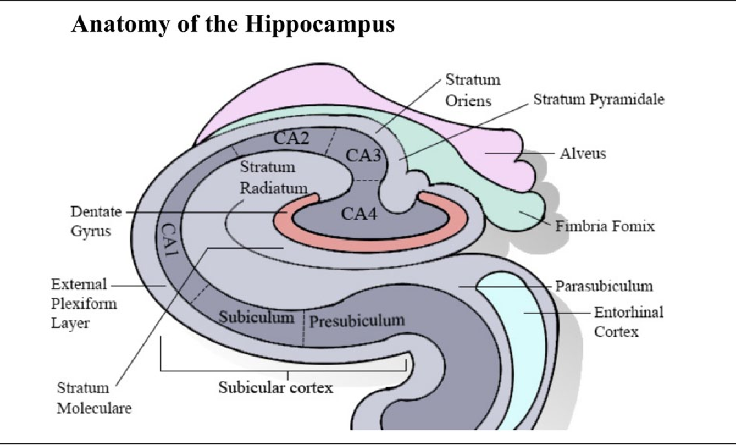
Image Source: Ammon's Horn 2 (CA2) of the Hippocampus: A Long-Known Region with a New Potential Role in Neurodegeneration \(^{[3]}\)
Dentate Gyrus
To continue.
Appendix
Limbic System
The limbic system is a part of the primitive brain and its primary functions concern hunger, motivation, sex drive, mood, pain, pleasure, appetite, and memory.
Pyramidal Neurons
The pyramidal neuron, named after the conic-shaped soma, is a multipolar neuron and is the primary excitation unit of the mammalian prefrontal cortex and corticospinal tract. These neurons are also found in the hippocampus and amygdala. Studies on pyramidal neurons mainly include neuroplasticity and cognition studies.